It’s easy to feel confused when your doctor talks about MRI and CT scans. Are they just two versions of the same test, or do they offer different insights? These imaging techniques are both essential, but they work in unique ways.
MRIs create highly detailed images of soft tissues, while CT scans are faster and often used to diagnose bone and internal injuries. Understanding the differences can help you make the best choice for your care.
So, how do you decide whether MRI or CT scan is right for you?
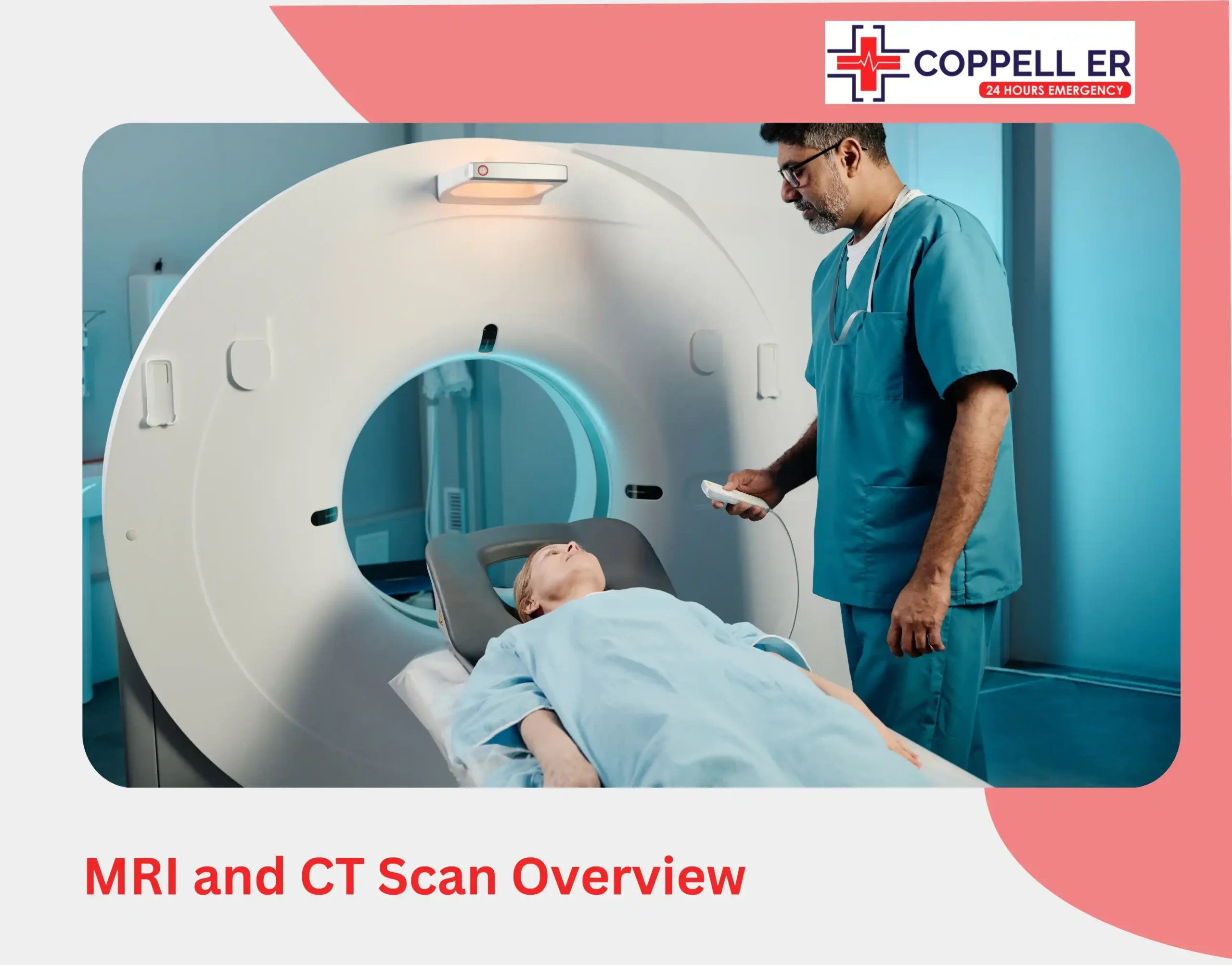
MRI and CT Scan Overview

Before diving into the specifics of brain imaging, it’s important to understand the basic principles behind MRI and CT scans.
- MRI (Magnetic Resonance Imaging)
MRI uses strong magnetic fields and radio waves to create precise images of organs, tissues, and structures in the body. Because it doesn’t use radiation, MRI is particularly useful for patients who need repeated scans or have specific health considerations. Its ability to distinguish between soft tissues make it valuable for neurological imaging, where detailed imaging is crucial.
- CT Scan (Computed Tomography)
A CT scan, also known as a CAT scan, uses X-rays to create cross-sectional images of the body. These images are combined to produce a detailed view of the internal structures. While CT scans expose patients to small amounts of radiation, they are quick and effective at visualizing bones, blood vessels, and internal injuries.
Key Differences Between MRI and CT for Brain Imaging
When comparing brain imaging techniques, there are several key factors to consider:
Image Detail and Quality
- MRI: Offers exceptional detail, especially for soft tissues such as the brain. It can show differences between gray matter, white matter, and cerebrospinal fluid, making it a superior choice for neurological imaging when precise details are required. MRI can also detect smaller lesions and abnormalities that a CT scan might miss.
- CT Scan: Provides excellent images of bones and hard structures, but its resolution for soft tissues like the brain is lower compared to MRI. However, it is still highly effective for detecting issues like bleeding, skull fractures, and acute injuries.
Speed of Imaging
- MRI: Generally takes longer, with scans lasting from 30 minutes to over an hour, depending on the area being scanned and the level of detail required. This makes MRI less suitable for emergency situations where rapid imaging is essential.
- CT Scan: Much faster, typically completed within 5 to 10 minutes. The quick turnaround time makes it a preferred option in emergency settings, such as trauma cases or stroke assessment, where speed is critical.
Radiation Exposure
- MRI: Does not use ionizing radiation, making it safer for repeated use, especially in patients who need multiple scans or in populations like pregnant women and children.
- CT Scan: Uses X-rays, which expose the patient to a small amount of radiation. While the levels are generally considered safe, repeated exposure can increase long-term risks, making CT less ideal for ongoing monitoring or in sensitive populations.
Ability to Image Acute Conditions
- MRI: While MRI is excellent for identifying soft tissue abnormalities, it is not typically the first choice for acute conditions like trauma or stroke due to the longer scan time. However, it is valuable for assessing ongoing issues such as tumors, multiple sclerosis, or brain inflammation.
- CT Scan: The go-to option for acute brain injuries, including stroke, hemorrhage, and skull fractures. The rapid imaging makes it ideal for emergency room settings.
Patient Comfort and Considerations
- MRI: Some patients find the MRI experience uncomfortable due to the enclosed space and the loud noises generated by the machine. Additionally, people with certain metal implants, such as pacemakers, may not be eligible for MRI due to the magnetic field.
- CT Scan: Generally more comfortable for patients because it’s quicker and less noisy. Since it doesn’t rely on a strong magnetic field, people with most types of implants can safely undergo a CT scan.
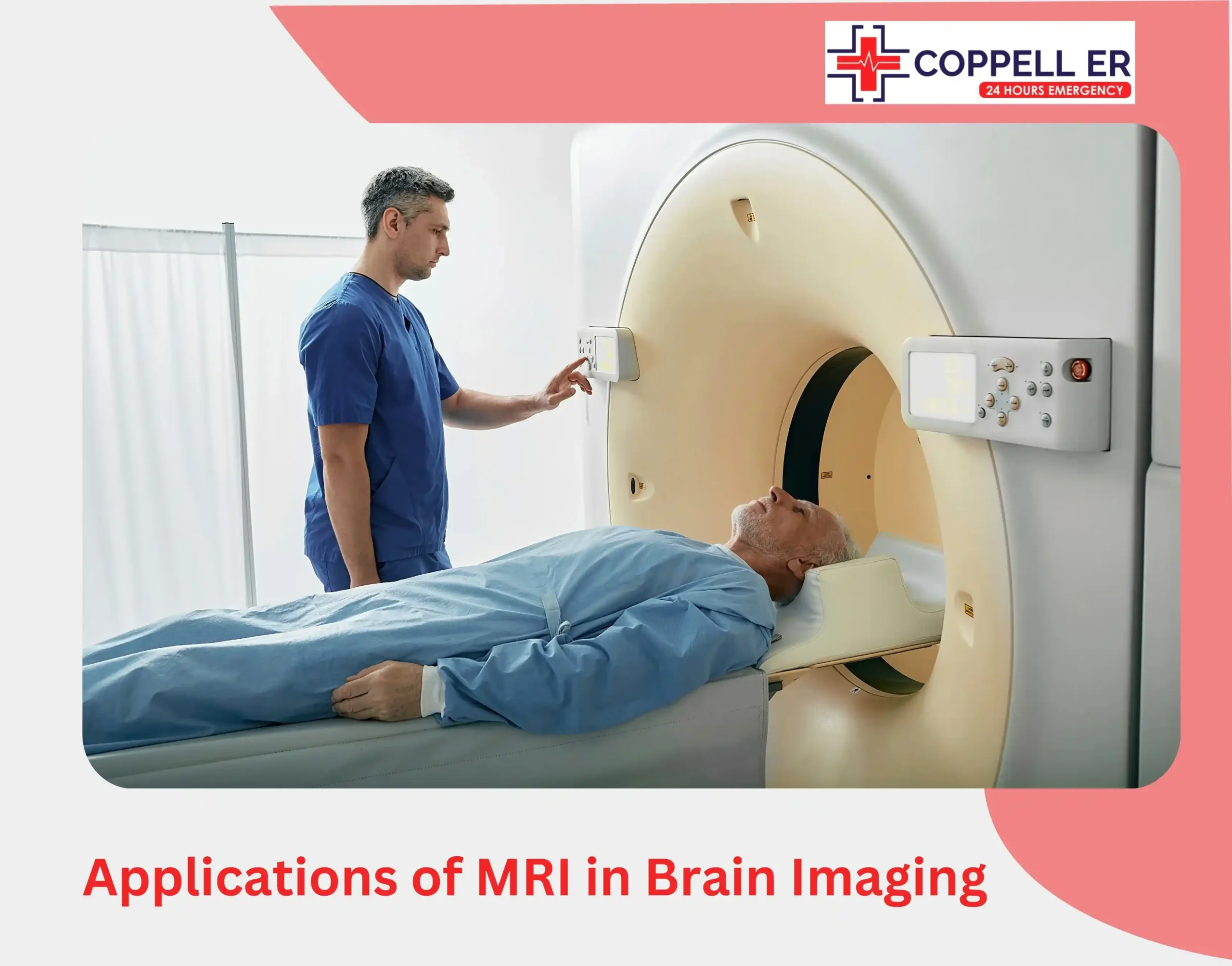
Applications of MRI in Brain Imaging

MRI is often the first choice when detailed brain imaging is needed for diagnosis and treatment planning. Some of the common uses of MRI in neurological imaging include:
Neurological Disorders
MRI is frequently used to diagnose and monitor neurological conditions like multiple sclerosis (MS), where it can detect lesions in the brain and spinal cord. MRI is highly sensitive in identifying changes in white matter, which is critical in MS management.
Brain Inflammation
MRI is an excellent tool for identifying inflammation in brain tissues, such as in conditions like encephalitis or meningitis. Its ability to differentiate between different types of soft tissue makes it invaluable in detecting areas of inflammation or infection.
Neurodegenerative Diseases
For conditions like Alzheimer’s disease, Parkinson’s disease, or Huntington’s disease, MRI plays a crucial role in identifying early signs of brain atrophy and structural changes that correlate with these disorders.
Functional MRI (fMRI)
fMRI is a specialized type of MRI that measures and maps brain activity by detecting changes in blood flow. It’s particularly useful for research and pre-surgical planning, especially in cases involving brain mapping for cognitive functions.
Applications of CT Scan in Brain Imaging
CT scans are often preferred for urgent medical situations and cases where rapid diagnosis is necessary. Key uses of CT for neurological imaging include:
Stroke and Hemorrhage
CT is highly effective in detecting intracranial hemorrhage and identifying blockages or bleeding in the brain that indicate a stroke. In emergency settings, CT is the preferred method to quickly assess whether a patient is experiencing an ischemic or hemorrhagic stroke.
Head Trauma
After accidents or head injuries, a CT scan is usually the first imaging test conducted. It’s excellent for identifying skull fractures, brain bleeding, and other immediate concerns related to trauma.
Hydrocephalus
CT can quickly detect hydrocephalus, a condition where there is an abnormal buildup of cerebrospinal fluid in the brain, leading to increased pressure. The fast imaging capabilities allow for quick decisions in managing the condition.
Brain Aneurysms
Although MRI is useful in detecting aneurysms, a CT angiography (CTA), which involves the use of contrast material, is particularly effective for identifying aneurysms and abnormalities in the blood vessels of the brain.
Infections and Abscesses
CT scans can help detect brain infections and abscesses, particularly when quick imaging is needed to determine the severity and spread of the infection.
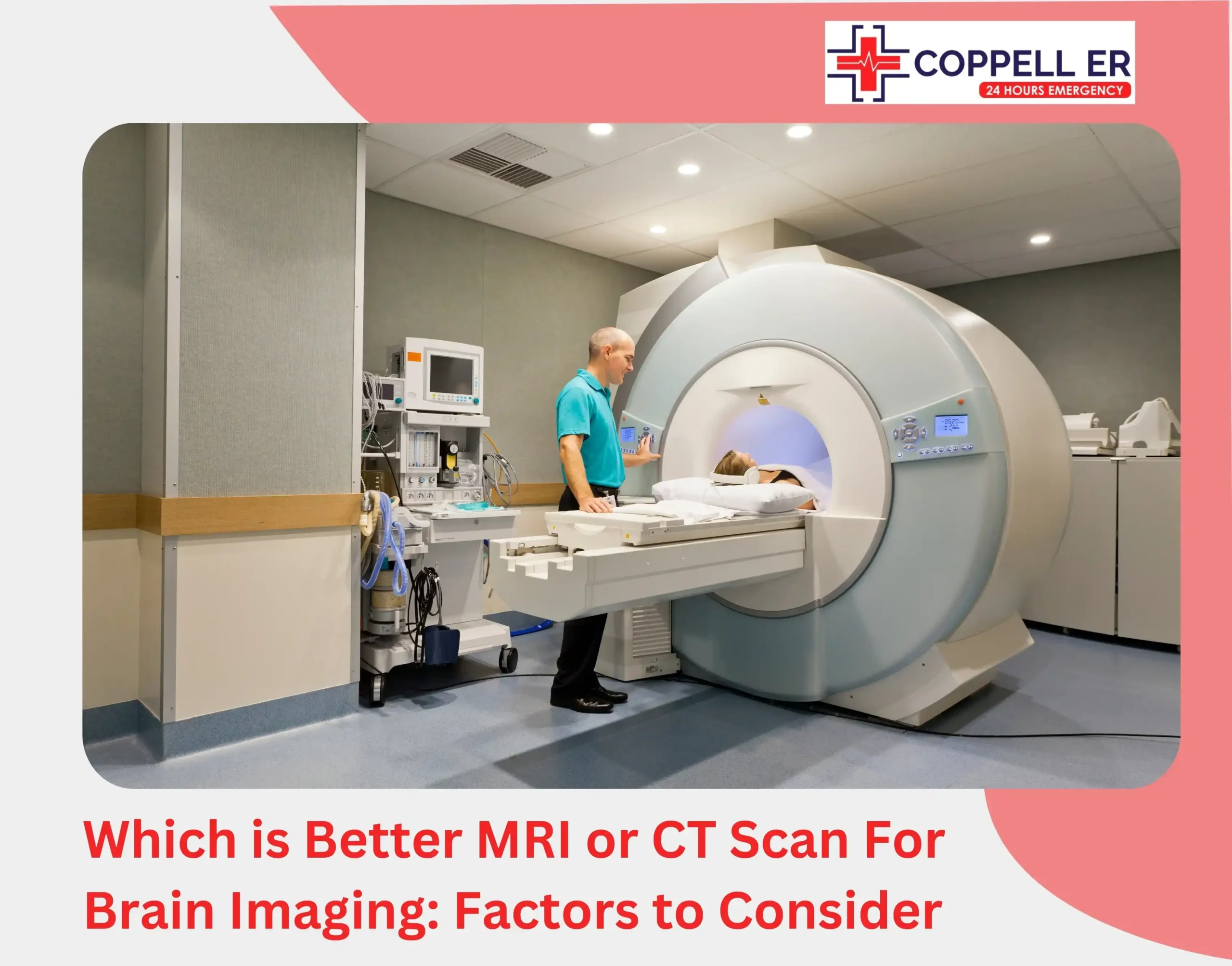
Which is Better MRI or CT Scan For Brain Imaging: Factors to Consider

When deciding whether to use brain CAT scan vs MRI, several factors come into play:
Condition Suspected
For conditions like tumors, neurodegenerative diseases, or inflammation, MRI is usually the better choice due to its superior soft tissue contrast. However, for acute issues such as trauma or stroke, a CT scan is often the first imaging modality used.
Urgency
In cases where time is of the essence, such as emergency room scenarios, CT scans are preferred due to their speed. MRI, while providing more detailed images, takes longer and may not be suitable in time-critical situations.
Radiation Exposure
For patients who require repeated imaging, such as those with chronic conditions like epilepsy, seizures, or multiple sclerosis, MRI is preferable due to the absence of radiation. CT, while useful, exposes the patient to ionizing radiation, which can accumulate over time.
Patient’s Physical Condition
Patients with metal implants or those who are claustrophobic may not be good candidates for MRI, which requires patients to lie still in an enclosed tube. In these cases, CT may be the safer or more comfortable option.
Key Takeaway
There isn’t a one-size-fits-all answer to whether an MRI or CT scan is better for brain imaging. In emergencies like trauma, stroke, or sudden neurological changes, a CT scan is typically the first choice due to its speed and ability to detect acute injuries or bleeding quickly. However, for chronic conditions such as tumors or detailed neurological evaluations, MRI is preferred for its superior imaging quality.
When comparing MRI vs X-Ray vs CT Scan, it’s important to note that X-rays are seldom used for brain imaging, while MRI and CT scans are essential for diagnosing brain-related issues.
ER of Coppell offers quick access to emergency CT scan, providing quick access to advanced CT scans when you need immediate answers.We also provide comprehensive laboratory services and appropriate referrals for MRI scans when needed.
[Visit Coppell ER for CT Imaging Today]
FAQs
Is CT or MRI better for headaches?
For most headaches, CT scans are used to rule out emergencies like bleeding, while MRI is better for diagnosing underlying causes such as tumors or nerve issues.
Does MRI or CT scan show brain tumor?
MRI is generally better for detecting brain tumors due to its detailed images of soft tissues, while CT scans can also identify tumors but are less detailed.
When to Use CT vs MRI scan?
CT Scans are preferred in emergencies like head trauma or suspected bleeding due to their speed and rapid results. MRIs are typically chosen for detailed examination of tumors and nerve conditions when time isn’t critical.
Can MRI results be seen immediately?
MRI results typically cannot be seen immediately. Images are processed and analyzed by a radiologist, and results are usually available within a few days.


