
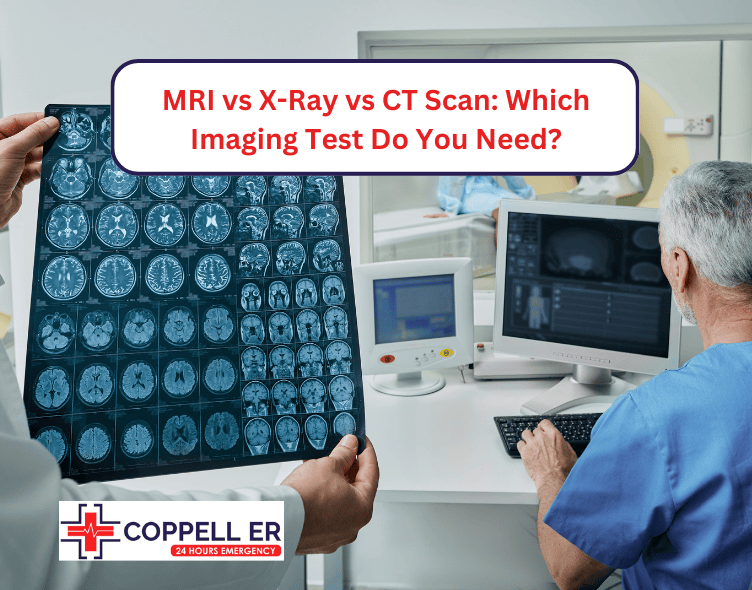
When injuries occur or unexplained symptoms persist, medical imaging becomes crucial. MRI, X-ray, and CT scan each serve as powerful diagnostic tools, offering unique insights into the human body.
From detailed soft tissue analysis to rapid skeletal assessments, these three types of imaging techniques empower medical professionals to see beneath the surface with remarkable precision. But what sets each technique apart, and how do doctors determine the most suitable option?
Whether you’re dealing with a sports injury or managing a chronic condition, understanding these tools enables you to actively participate in your care journey.
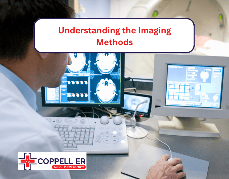
Understanding the Imaging Methods
MRI (Magnetic Resonance Imaging)
An MRI uses strong magnetic fields and radio waves to produce detailed images of the organs, tissues, and other internal structures of the body. Unlike X-rays and CT scans, an MRI does not use ionizing radiation, which makes it safer for certain patients.
How MRI Works
In an MRI scan, the patient lies on a table that slides into a large tube surrounded by magnets. The magnetic field aligns hydrogen atoms in the body, and radio waves are then used to disturb this alignment. As the atoms return to their original positions, they emit signals that are captured by the MRI machine and converted into highly detailed images by a computer.
When MRI Is Required
MRI is often used to examine soft tissues in great detail, making it particularly useful for diagnosing conditions in the brain, spinal cord, joints, muscles, and ligaments. Common cases where MRI is required include:
- Diagnosing brain disorders (e.g., tumors, strokes, or multiple sclerosis)
- Identifying issues with the spinal cord (e.g., herniated discs or spinal injuries)
- Assessing joint or soft tissue injuries
- Diagnosing cardiovascular problems (heart or blood vessels)
- Evaluating the extent of cancer
X-Ray
An X-ray is one of the oldest and most widely used imaging methods. It involves the use of electromagnetic radiation to produce images of the inside of the body. X-rays are particularly good at showing bones and are often used to detect fractures or abnormalities in the skeletal structure.
How X-ray Works
X-rays work by passing a small amount of ionizing radiation through the body. Different tissues absorb this radiation in varying amounts: dense tissues like bones absorb more X-rays and appear white on the image, while softer tissues absorb less and appear in shades of gray. Air-filled spaces, like the lungs, appear black.
When X-ray Is Required
X-rays are required in a variety of situations, most commonly to:
- Diagnose fractures or bone dislocations
- Detect infections or fluid in the lungs (e.g., pneumonia)
- Evaluate joint degeneration or arthritis
- Identify dental issues (e.g., cavities or tooth infections)
- Locate foreign objects in the body
X-rays are generally the first imaging option due to their simplicity and efficiency, particularly in emergency situations.
CT Scan (Computed Tomography)
A CT scan, or CAT scan, combines X-rays and computer technology to create detailed cross-sectional images of the body. It provides more detailed information than a standard X-ray and is particularly useful for visualizing bones, blood vessels, and soft tissues all in one scan.
How CT Scan Works
During a CT scan, the patient lies on a table that moves through a circular machine that houses an X-ray tube and detectors. The X-ray tube rotates around the body, sending X-rays through the body from multiple angles. A computer processes these images to generate cross-sectional slices of the body, which can then be viewed as a three-dimensional image.
When CT Scan Is Required
A CT scan is required when doctors need detailed information quickly, especially in emergencies. Some common uses include:
- Diagnosing traumatic injuries (e.g., head, chest, or abdominal injuries)
- Detecting internal bleeding
- Identifying cancers or tumors
- Evaluating bone fractures or spine injuries in greater detail than an X-ray
- Planning for surgeries (e.g., to determine the exact location of a tumor)
MRI vs X-ray vs CT Scan: Imaging Procedure Differences
Each of these imaging methods is distinct in terms of the procedure, the technology used, and the quality of the images produced. Understanding these imaging procedure differences is key to knowing which method is best suited for a particular medical condition.
Image Detail and Resolution
- MRI: Of the three methods, an MRI provides the most detailed images, particularly for soft tissues. It offers a high level of contrast between different tissue types, making it the best choice for visualizing the brain, muscles, ligaments, and tendons. For example, if a patient has a suspected ligament injury, an MRI would likely be the preferred imaging method.
- X-ray: X-rays are best for capturing images of bones and can quickly identify fractures or joint dislocations. However, they are not as useful for soft tissue evaluation since the image quality for non-bone structures is limited.
- CT Scan: A CT scan offers a middle ground between MRI and X-ray. It provides detailed images of both bones and soft tissues, though it may not be as precise as an MRI for soft tissue visualization. CT scans are particularly effective for detecting internal bleeding, fractures, and tumors, and they are faster than MRIs, making them ideal for emergency diagnostics.
Time and Efficiency
- MRI: MRI scans are often time-consuming, lasting anywhere from 30 minutes to over an hour, depending on the area being scanned. The patient must remain very still to avoid image distortion, which can be challenging for some individuals.
- X-ray: X-rays are extremely fast, often taking only a few minutes to complete. This makes them ideal for emergency situations where time is of the essence.
- CT Scan: CT scans typically take only a few minutes, though the preparation time can be longer if a contrast dye is needed. This efficiency makes CT scans particularly useful in urgent medical situations.
Radiation Exposure
- MRI: One of the major advantages of MRI is that it does not use ionizing radiation, which makes it safer for certain populations, including pregnant women and children, who are more vulnerable to the harmful effects of radiation.
- X-ray: X-rays do involve exposure to ionizing radiation, though the levels are generally considered low and safe for most patients. However, repeated exposure to X-rays can increase the risk of cancer, so they are used sparingly, particularly in children.
- CT Scan: CT scans involve significantly more radiation than a standard X-ray because they require multiple X-ray images to create cross-sectional slices of the body. As such, doctors weigh the benefits and risks carefully before recommending a CT scan, particularly for children and pregnant women.
Which is Better: MRI, X-ray, or CT Scan?

The choice between MRI, X-ray, or CT scan depends on the patient’s specific condition and the area of the body being examined. Each imaging method has its strengths and weaknesses:
- MRI is the best option for soft tissue evaluation and is preferred when doctors need detailed images of the brain, spinal cord, joints, or muscles. It is also safer for patients who should avoid radiation.
- X-rays are fast, cost-effective, and ideal for diagnosing bone fractures, lung infections, and joint problems. They are often the first imaging test used in emergency situations.
- CT scans are highly detailed and provide a balance between bone and soft tissue imaging, making them ideal for trauma cases, internal bleeding detection, and tumor evaluations.
Ultimately, the decision to use an MRI, X-ray, or CT scan is guided by the patient’s condition, the body part in question, and the level of detail required for diagnosis. In some cases, doctors may recommend a combination of these imaging techniques for a more comprehensive evaluation.
At our facility, we house a certified full-service laboratory Coppell offering accurate diagnoses through state-of-the-art imaging. From routine X-rays to specialized CT scanning services Coppell, our facility is equipped to meet all your diagnostic imaging needs under one roof.
Schedule Your Imaging Consultation
FAQs
Why Do Doctors Not Recommend MRI?
Doctors may not recommend MRI due to high costs, longer scan times, and issues with claustrophobia or metal implants.
Which Is More Radiation, CT or MRI?
CT scans expose patients to more radiation than MRI scans. CT scans use X-rays to create detailed images, while MRIs do not use ionizing radiation at all.
What Is the Most Expensive Scan?
MRI scans are generally the most expensive imaging option compared to X-rays and CT scans. The costs can vary based on factors like location, facility, and the specific type of MRI being performed.