
Traditional X-rays take 15-30 minutes to develop in a darkroom. Digital X-rays appear on screen in seconds. In emergency medicine, this difference directly impacts patient outcomes and treatment decisions.
The gap between digital X-rays vs normal X-rays extends far beyond speed. Image quality, radiation exposure, and diagnostic accuracy all favor digital technology. Yet many patients don’t understand why this matters when they need emergency care.
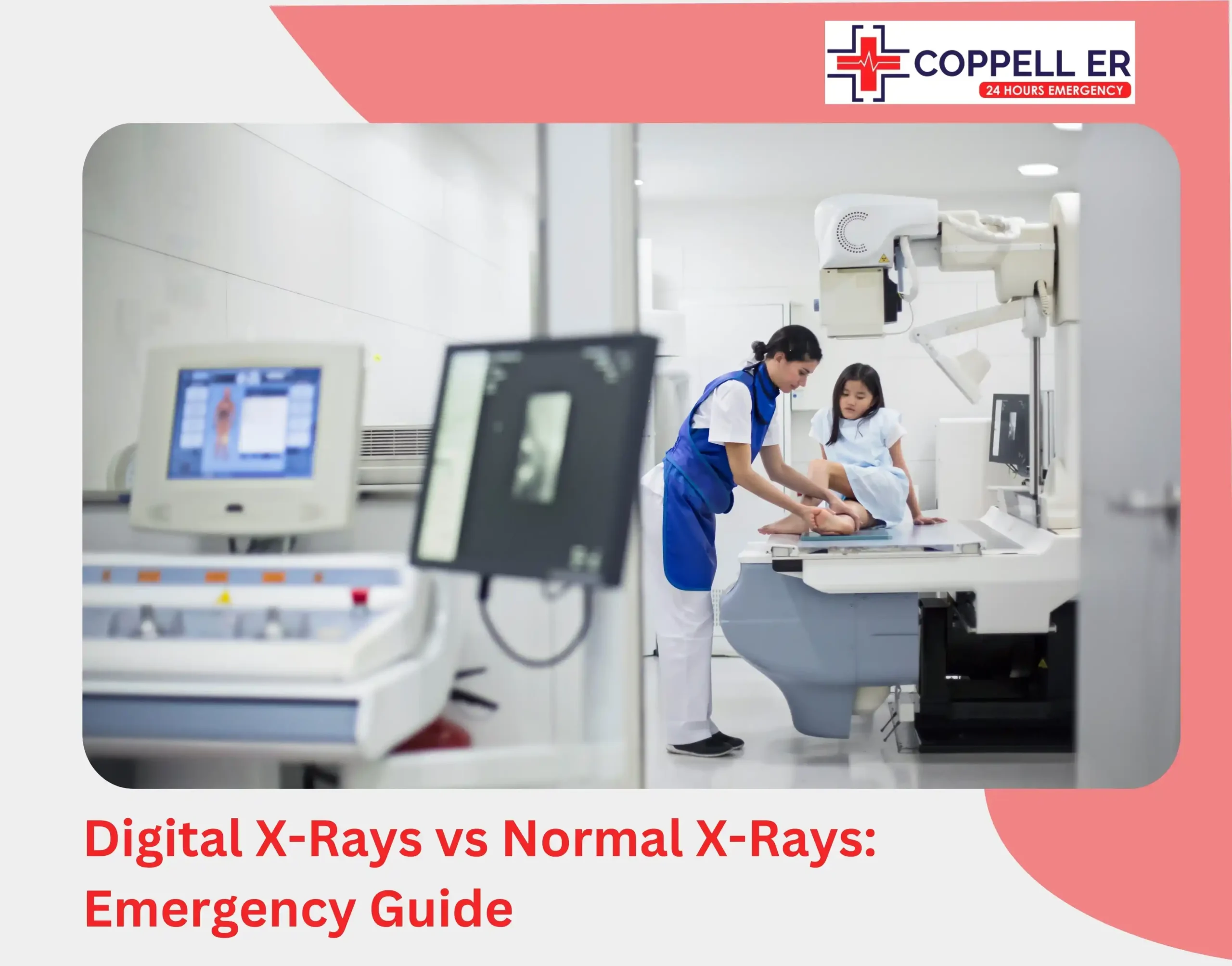
Here’s exactly how this technology improves emergency treatment at Coppell ER.
How Is Digital X-Ray Better Than a Traditional X-Ray?

Traditional X-rays used photographic film that had to be developed in a darkroom using chemicals. This process could take anywhere from 10–30 minutes or more.
Once developed, the film had to be dried, physically handled and passed around for sharing with other departments. All films had to be stored in physical files. If someone needed a past image, it meant manually digging it out.
Digital X-rays use electronic sensors to capture images of the inside of the body. It’s like switching from an old disposable camera to a smartphone. These digital images are instantly available on computers and can be shared with specialists at other locations in seconds.
4 Critical Advantages of Digital X-Rays Over Traditional X-Rays
1. Speed That Saves Lives
Digital X-rays provide instant radiology results compared to traditional film processing that takes 15-30 minutes. This speed advantage directly impacts treatment decisions, whether diagnosing a collapsed lung, internal bleeding, or spinal fracture, immediate imaging allows emergency physicians to begin appropriate care without delay.
2. Image Clarity For Better Diagnosis
While speed is essential, clarity is equally critical in making an accurate diagnosis. Traditional X-rays often struggle to capture subtle fractures, small dislocations, soft tissue damage, or early signs of infection. With digital X-Rays, clinicians gain exceptional control over image manipulation, allowing them to:
- Zoom into specific areas to detect hidden injuries
- Enhance contrast for better visualization of soft tissues
- Compare prior images side by side, tracking progress over time
For instance, a patient with wrist pain after a fall may have a minor stress fracture that film X-rays could easily miss. Digital X-ray shows a clear image of even the faintest fractures. Early diagnosis and treatment means no long-term complications like joint instability or chronic pain.
3. Seamless Sharing Between Teams
Digital x-rays make it super easy to share images with everyone who needs to see them, whether they’re at another hospital across town. Traditional X-rays required physical film transport, adding critical time to emergency transfers.
For critical conditions like spinal injuries or internal bleeding, having your diagnostic images readily available means treatment can begin immediately upon transfer.
4. Reduced Radiation Exposure
While efficiency and precision are at the heart of digital x-rays, patient safety remains a top priority. Film-based X-rays require significantly higher radiation exposure, posing long-term health risks. It is riskier for patients requiring frequent imaging, such as pregnant women and children.
Digital X-rays can reduce radiation exposure by up to 80%.1 This matters even more in the ER where patients may need multiple scans. Also, it’s ideal for vulnerable populations like children, pregnant women, or the elderly.
How Digital X-rays Coppell ER Support Different Emergencies

Let’s take a look at how Coppell ER uses digital X-rays to handle a variety of emergencies:
- Trauma Care: In motor vehicle accidents, digital X-Rays Coppell ER help identify internal fractures, bone displacement, or rib injuries within minutes. This guides decisions on stabilization, transfer, or surgical needs.
- Pulmonary Emergencies: In patients with shortness of breath, digital imaging Coppell ER can quickly reveal pneumonia, pneumothorax, or pleural effusion. These insights guide immediate respiratory support or drainage procedures.
- Foreign Object Detection: Whether it’s a child swallowing a coin or an adult with suspected shrapnel injury, digital x-rays offer precise localization that is essential for surgical retrieval.
Digital X-Rays vs Normal X-Rays: The Bottom Line

The differences between digital X-rays vs normal X-rays become critical in emergency situations. Digital technology delivers faster results, clearer images, reduced radiation, and seamless information sharing. In emergency settings, this directly impacts patient care quality and recovery outcomes.
When you need emergency imaging, a digital x-ray isn’t just a convenience.Its an essential tool that helps emergency physicians make faster and more accurate treatment decisions.
FAQs
Are digital X-rays safer than traditional X-rays?
Yes! Digital X-rays use lower doses of radiation, making them a safer imaging option, especially for patients who require multiple scans.
How long does it take to get results from a digital X-ray in an emergency setting?
Images are available within seconds, allowing medical teams to assess injuries or conditions almost immediately.
Can digital X-rays detect all types of injuries and conditions?
While they are excellent for diagnosing fractures, infections, and certain internal injuries, additional imaging like CT scans or MRIs may be needed for more complex cases.
Can children and pregnant women safely undergo digital X-rays?
Yes, but precautions are taken to minimize radiation exposure. Lead shielding may be used, and alternative imaging methods may be considered if necessary.
How do digital X-rays help diagnose fractures in emergency settings?
Digital X-Rays provide high-resolution images that reveal bone fractures, allowing doctors to quickly assess the severity and location of the break. This advanced fracture diagnosis capability enables emergency teams to make rapid and accurate treatment decisions for best possible patient outcomes.